| |
| Step1 |
|
|
|
|
Case
ID: |
063099.18 |
| Body Weight: |
19 g |
|
|
| Procedures: |
|
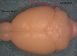
The dorsal view of a fixed mouse brain. |
|
| Step2 |
|
|
|
|
Case
ID: |
063099.18 |
| Body Weight: |
19 g |
|
|
| Procedures: |
|
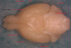
The ventral view of the brain. From this view you can see the
chiasm. By convention the optic nerve is the fiber form eyeball to
chiasm, the fiber arise from ganglion cells in the retina. From
chiasm to its end in the lateral geniculate is called optic tract. |
|
| Step3 |
|
|
|
|
Case
ID: |
063099.18 |
| Body Weight: |
19g |
|
|
| Procedures: |
|
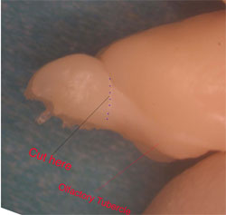
The temporal view of the brain. From this angle you can see there is
a separate line between the OB and the rest of the forebrain. Cut
here vertically to separate the bulbs. Typical weights of adult OB
are --- mg (bilateral fixed and ---- mg fresh) |
|
| Step4 |
|
|
|
|
Case
ID: |
063099.18 |
| Body Weight: |
19g |
|
|
| Procedures: |
|
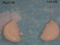
Bulbs are separated. This is a medial view. |
|
|
 |

|
|