Use the image below to select a section through the brain. Click on a button to view the corresponding section, or use the numbered guide in the frame below. Scroll down for more background on this atlas.
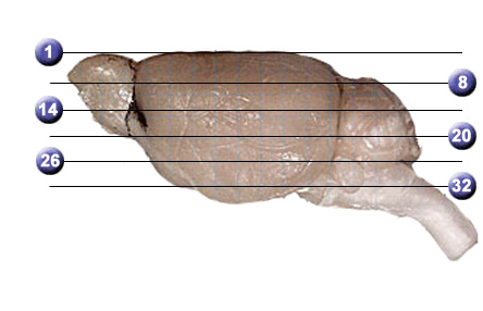
The markers above are spaced 1 mm apart and correspond to levels of the Atlas. The zero coordinate is the bregma point, a landmark visible on the skull. To use this atlas effectively you may need to assign your browser more memory than usual. If you are connecting at 56K you will also need patience. Many of the jpeg-compressed images are 100K to 500K.
If you click on the buttons for "grid" or "labels" in the brain section area before the page finishes loading images, you may not be able to view the gridded or labeled images without reloading the page. JavaScript must be enabled to view the small versions of the gridded and labeled sections, but the enlarged images can be viewed with any settings. They are so large that you will need to use the scroll bars to view different parts of the image. You may want to copy pages of the Altas to your computer and then view them using a program such as Photoshop.
About this DBA/2J Horizontal Atlas: The brain is that of a 203-day-old DBA/2J male with a body weight of 27 grams and a brain weight (fixed) of 397 milligrams. In vivo brain weight was probably about 405–410 mg. The animal's celloidin case ID number is 247 and you can view one of the two slides used to make this atlas. Shrinkage during fixation and processing is 24% (linear). The in vivo grid compensates for this shrinkage. The abbreviations we have used to label the sections conform to those in the Franklin-Paxinos atlas (The Mouse Brain in Stereotaxic Coordinates, Academic Press, San Diego, 1998, ISBN Number 0-12-266070-6).
The brain and sections were all processed as described in our methods section. The enlarged images have a pixel count of 3056 x 2032 and the resolution is 3.5 microns/pixel for the processed sections. All images are 8 bits per RBGB channel JPEG images.
Plans: In the next several years we hope to add several additional atlases of the same sort for other strains of mice. A horizontal
C57BL/6J atlas and a DBA/2J coronal atlas were completed by Tony Capra, summer 2000. This high resolution color atlas was completed by Aida Causevic, July 2001. We plan to produce additional atlases over the next several years. As described in our MBL Procedures Section, it is not hard to make your own strain-specific atlas from the high resolution images in the MBL.