| |
| Step1 |
|
|
|
|
Strain
ID: |
DBA/2J |
| Body Weight: |
21 g |
|
|
| Procedures: |
|
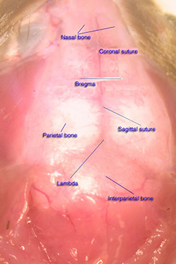
This is a dorsal view of a mouse's skull, in order to access the
brain you can cut along the coronal suture and sagittal suture then
pull off both sides of parietal bone and interparietal bone |
|
| Step2 |
|
|
|
|
Strain
ID: |
DBA/2J |
| Body Weight: |
21 g |
|
|
| Procedures: |
|
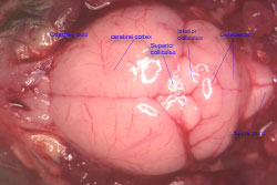
Now the skull is open and you can see the brain clearly, in order to
get the hippocampus you'd better keep the brain in the cranial
cavity so the brain will not move when you continue with the
following steps |
|
| Step3 |
|
|
|
|
Strain
ID: |
DBA/2J |
| Body Weight: |
21g |
|
|
| Procedures: |
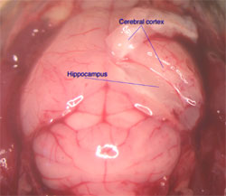
In order to expose the hippocampus you need to remove the cerebral
cortex covering it. The first incision is at the end of the
hemisphere; the incision should be about 0.7mm deep for most adult
mouse that you might not hurt the hippocampus while to expose it.
The 2nd incision is about 1.5-2mm in front of the first one, this
incision you need cut into the lateral ventricle, both of the
incisions go to the ventral of the brain and meet there. Now this
piece of cortex is free, pull it up, you will see the hippocampus
just like in this picture, also you can see the CSF in the opened
ventricle.
|
|
| Step4 |
|
|
|
|
Strain
ID: |
DBA/2J |
| Body Weight: |
21g |
|
|
| Procedures: |
|
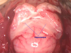
Keep working on the other side of the brain pull up both sides of
the cortex that covering the hippocampus along the ventricle. Now
you can see the dorsal part of the hippocampus. Separate the rest of
the hippocampus from the cortex covering it along the surface of the
hippocampus towards the ventral part of the hippocampus. |
|
|
 |

|
|