Use the image below to select a section through the brain. The bregma
zero coordinate is marked by the grey circle. Click on a button to view the
corresponding section, or use the numbered guide in the frame below. Scroll down
for more background on this atlas.
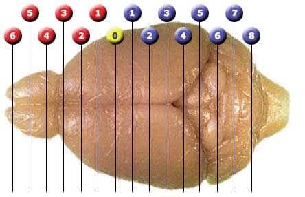
The markers above correspond to levels of the Atlas. The zero coordinate is the bregma
point, a landmark visible on the skull. To use this atlas effectively you may need to assign
your browser more memory than usual. If you are connecting at 56K you will also
need patience. Many of the jpeg-compressed images are 100K to 500K.
If you click on the buttons for "grid" or "labels" in the brain section area
before the page finishes loading images, you may not be able to view the gridded
or labeled images without reloading the page. JavaScript must be enabled to view
the small versions of the gridded and labeled sections, but the enlarged images
can be viewed with any settings. They are so large that you will need to use the
scroll bars to view different parts of the image. You may want to copy pages of
the Altas to your computer and then view them using a program such as Photoshop.
About this Atlas: The brain is that of a 76-day-old DBA/2J female with a body
weight of 20.7 gm and a brain weight (fixed) of 401.2 mg. The animal's celloidin
case ID number is 165 and you can view the two slides (a, b) used to make this atlas. Shrinkage during fixation and
processing is 21% (linear). The in vivo grid compensates for this shrinkage. The
anterior-posterior coordinates are taken from an excellent print atlas of a
C57BL/6J brain by K. Franklin and G. Paxinos (The Mouse Brain in Stereotaxic
Coordinates, Academic Press, San Diego, 1997, ISBN Number 0-12-26607-6; Library
of Congress: QL937.F72). The abbreviations we have used to label the sections conform to
those in the Franklin-Paxinos atlas.
A C57BL/6J mouse brain may contain as many as 75 million neurons, 23 million
glial cells, 7 million endothelial cells associated with blood vessels, and 3 to
4 million miscellaneous pial, ependymal, and choroid plexus cells (see data
analysis in Williams, 2000). We have not yet counted total cell number in DBA/2J mice, but the counts are probably appreciably lower.
The brain and sections were all processed as described in our methods section. The enlarged images have a pixel count of 1865
x 1400 and the resolution is 4.5 microns/pixel for the processed sections.
Plans: In the next several years we hope to add several additional atlases of the same sort for
other strains of mice. A horizontal
C57BL/6J atlas and a DBA/2J coronal atlas were completed by Tony Capra, summer 2000, and additional atlases may
be made over the next several years. As describe in the MBL Procedures Section is not hard to make your own strain-specific atlas from the high resolution images in the MBL.